Bone Cross Section Microscope : Cross Section Cut Of Liver Cirrhosis Under The Microscope ... : This simply involves placing a section of the bone on the microscope stage and viewing.
Bone Cross Section Microscope : Cross Section Cut Of Liver Cirrhosis Under The Microscope ... : This simply involves placing a section of the bone on the microscope stage and viewing.. We obtained 24 axial slices of the normal brain. A cross section of a human long bone. An mri was performed on a healthy subject, with several acquisitions with different weightings: Find the perfect human bone cross section stock photo. Your bone cross section stock images are ready.
The lining of the trachea this model shows a cross section of compact bone. Bone is an architecturally complex system that constantly undergoes structural and functional optimisation through renewal and repair. Cross section through middle metacarpal bones of vector. This simply involves placing a section of the bone on the microscope stage and viewing. To download this image, create an account.
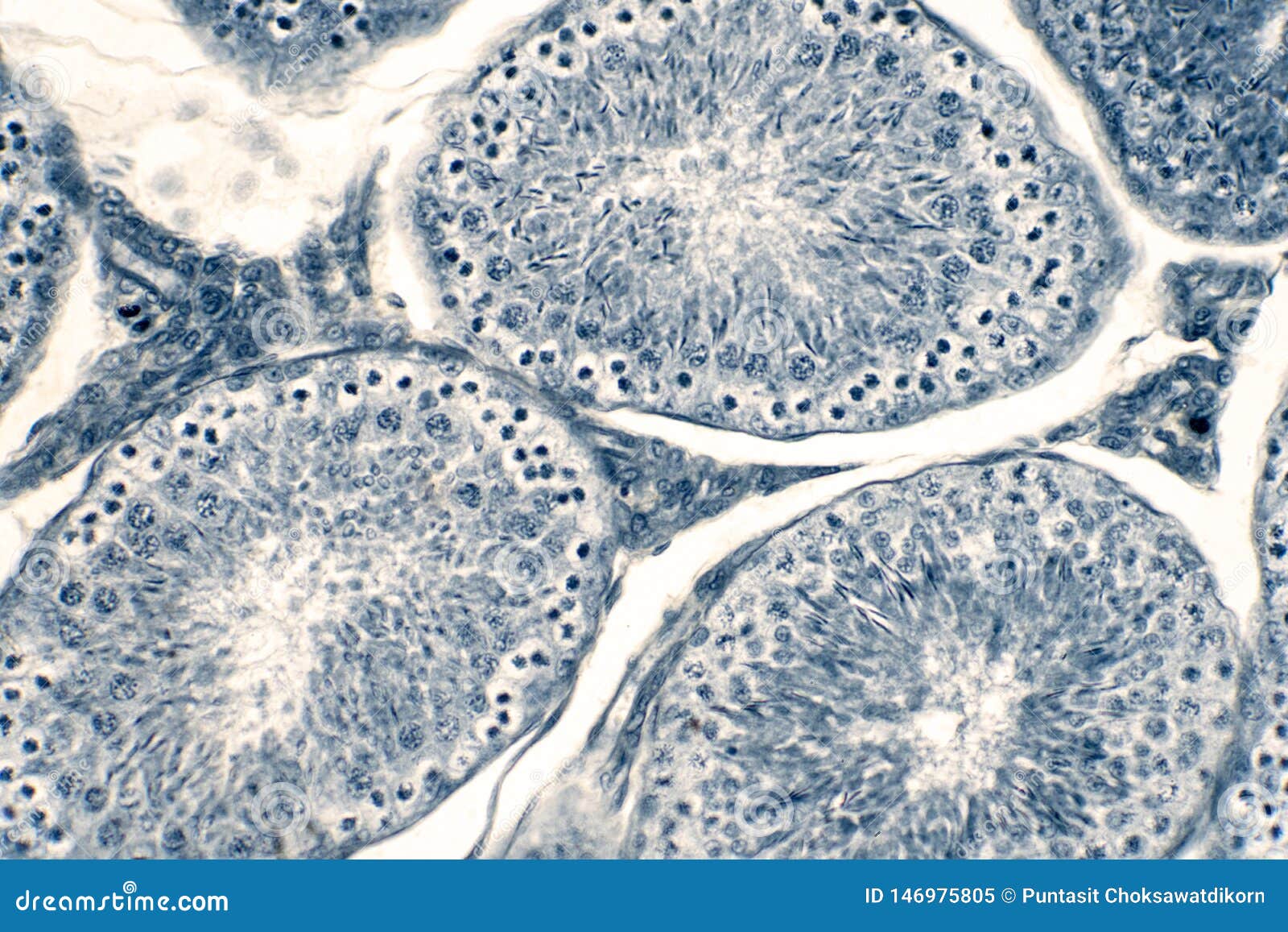
Cross Section Human Testis Under Microscope View Stock ... from thumbs.dreamstime.com Dreamstime is the world`s largest stock photography community. Spinal cord is one of the major structures of the body that helps in transmitting neuronal impulses from brain to rest of the body and vice versa. Thin section of dinosaur bone. Find the perfect human bone cross section stock photo. Coloured scanning electron micrograph (sem) of a section through an osteoclast bone cell in reabsorbing bone matrix, showing the cell's nucleus (round, centre). Both types of bone marrow are enriched with blood vessels and capillaries. The microscopic bone cross section image acquired by using electronic microscope and is shown in fig.2. Medically reviewed by the healthline medical network — written by the healthline editorial team — updated on january 20, 2018.
Observe that the matrix of the bone is deposited in concentric layers that are called lamellae.
Cross section of a bone, this image shows the interior of the bone, which has a lot of spongy bone tissue. From wikimedia commons, the free media repository. To download this image, create an account. Observe that the matrix of the bone is deposited in concentric layers that are called lamellae. Both types of bone marrow are enriched with blood vessels and capillaries. Back bone vertebrae thorax section in watercolor made from real life specimen, excellent for anatomical illustration anatomy article or. Thin section of dinosaur bone. The scanning electron microscope (sem) is among the most frequently used instruments for examining bone. Compact bone, spongy if you were to look at a piece of compact bone without the help of a microscope, it would seem to be. Coloured scanning electron micrograph (sem) of a section through an osteoclast bone cell in reabsorbing bone matrix, showing the cell's nucleus (round, centre). Compact bone cross section courtesy: Dreamstime is the world`s largest stock photography community. In a cross section of a bone you can usually see two types of bone tissue what are these called?
Find the perfect human bone cross section stock photo. Bone basics and bone anatomyhave you ever seen fossil remains of dinosaur and ancient human each bone in your body is made up of three main types of bone material: Bone marrow aspiration uses a hollow needle to remove a small sample (about 1 ml) of bone marrow for examination under a microscope. The microscopic bone cross section image acquired by using electronic microscope and is shown in fig.2. Cross section of a bone, this image shows the interior of the bone, which has a lot of spongy bone tissue.
Light microscopy micrographs at different magnifications ... from www.researchgate.net Your bone cross section stock images are ready. Bone marrow aspiration uses a hollow needle to remove a small sample (about 1 ml) of bone marrow for examination under a microscope. Which microscope is used to see the cross section of a stem? Use them in commercial designs under lifetime, perpetual & worldwide rights. Coloured scanning electron micrograph (sem) of a section through an osteoclast bone cell in reabsorbing bone matrix, showing the cell's nucleus (round, centre). Cross section through middle metacarpal bones of vector. Accuracy of the tested digitization method was expressed by. Cross section of a bone, this image shows the interior of the bone, which has a lot of spongy bone tissue.
Bone marrow aspiration uses a hollow needle to remove a small sample (about 1 ml) of bone marrow for examination under a microscope.
In a cross section of a bone you can usually see two types of bone tissue what are these called? The finished bone section will be bonded to a microscope slide and so the first step is to grind flat and polish the part of the bone that will be glued to the slide. The large dark spots are passages for blood if you were to look at it in under a microscope, it would look a lot like your kitchen sponge. Cross section through middle metacarpal bones of vector. This slide showing a cross section of the mammalian trachea (wind pipe) contains examples of several different kinds of tissues. Spinal cord is one of the major structures of the body that helps in transmitting neuronal impulses from brain to rest of the body and vice versa. Haversian systems comprise concentric rings of bone around a central channel or haversian canal. We obtained 24 axial slices of the normal brain. Compact bone cross section courtesy: Bones protect the various organs of the body, produce red and white blood cells, store minerals. From wikimedia commons, the free media repository. Compact bone, spongy if you were to look at a piece of compact bone without the help of a microscope, it would seem to be. Bone marrow aspiration uses a hollow needle to remove a small sample (about 1 ml) of bone marrow for examination under a microscope.
Coloured scanning electron micrograph (sem) of a section through an osteoclast bone cell in reabsorbing bone matrix, showing the cell's nucleus (round, centre). In a cross section of a bone you can usually see two types of bone tissue what are these called? Basic functions of bone bone is the basic unit of the human skeletal system and provides the framework for and bears the weight of the body, protects the vital organs, supports mechanical movement, hosts hematopoietic cells, and maintains iron homeostasis. The large dark spots are passages for blood if you were to look at it in under a microscope, it would look a lot like your kitchen sponge. Which microscope is used to see the cross section of a stem?
Pine Female Cone Cross Section Cut Under The Microscope ... from media.istockphoto.com The scanning electron microscope (sem) is among the most frequently used instruments for examining bone. This slide showing a cross section of the mammalian trachea (wind pipe) contains examples of several different kinds of tissues. Compact bone cross section courtesy: The lining of the trachea this model shows a cross section of compact bone. When the light that enters the condenser is polarized by placing a polarizer in the filter holder and a second, crossed polarizer at the image plane. Your bone cross section stock images are ready. Medically reviewed by the healthline medical network — written by the healthline editorial team — updated on january 20, 2018. Cross section through middle metacarpal bones of vector.
Bone cross section — stock image.
Both types of bone marrow are enriched with blood vessels and capillaries. Thin section of dinosaur bone. An mri was performed on a healthy subject, with several acquisitions with different weightings: The microscopic bone cross section image acquired by using electronic microscope and is shown in fig.2. Your bone cross section stock images are ready. Important features in the bone cross section such as harvesian canals, osteons, osteon fragments, lamellar bone, bony trabeculae, myxoid matrix and artifact for. Bones protect the various organs of the body, produce red and white blood cells, store minerals. Long and short bones, such as the femur and phalanges, arise from a cartilage model formed by endochondral ossification. Cross section of a bone, this image shows the interior of the bone, which has a lot of spongy bone tissue. Coloured scanning electron micrograph (sem) of a section through an osteoclast bone cell in reabsorbing bone matrix, showing the cell's nucleus (round, centre). Accuracy of the tested digitization method was expressed by. To download this image, create an account. This slide showing a cross section of the mammalian trachea (wind pipe) contains examples of several different kinds of tissues.
If your post declares something as fact bone cross section. Long and short bones, such as the femur and phalanges, arise from a cartilage model formed by endochondral ossification.